Michael Hooker Microscopy Facility (MHMF)
![]()
|
|
||
|
|
Michael Hooker Microscopy Facility (MHMF) |
|
|
|
||
Image Gallery (click on window to enlarge or play movies)
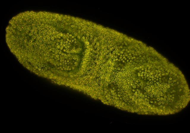 |
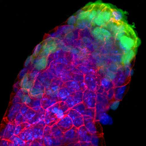
| Drosophila Embryo - Selective Plane Imaging Microscopy (R. Duronio Laboratory) |
| "Photoactivation" in the Alexa488 (green emission) channel after UV illumination used for DAPI imaging | |
 |
 |
| Before UV.tif | After UV.tif |
| 1st Captured Image. Note except for 3 cells most cells are dark in the green channel. | 3rd Captured Image - Note the increase in intensity of the nuclei in the green channel |
| Imaged widefield sequentially Ab1+TexasRed then Ab2+Alexa488 then DAPI. Single wavelength bandpass cubes. Images same gain and exposure times for each respective channel. | |
|
|
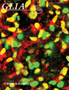
| On the cover of Glia - Jian Zou, Ryan Vetreno, Fulton Crews |
|
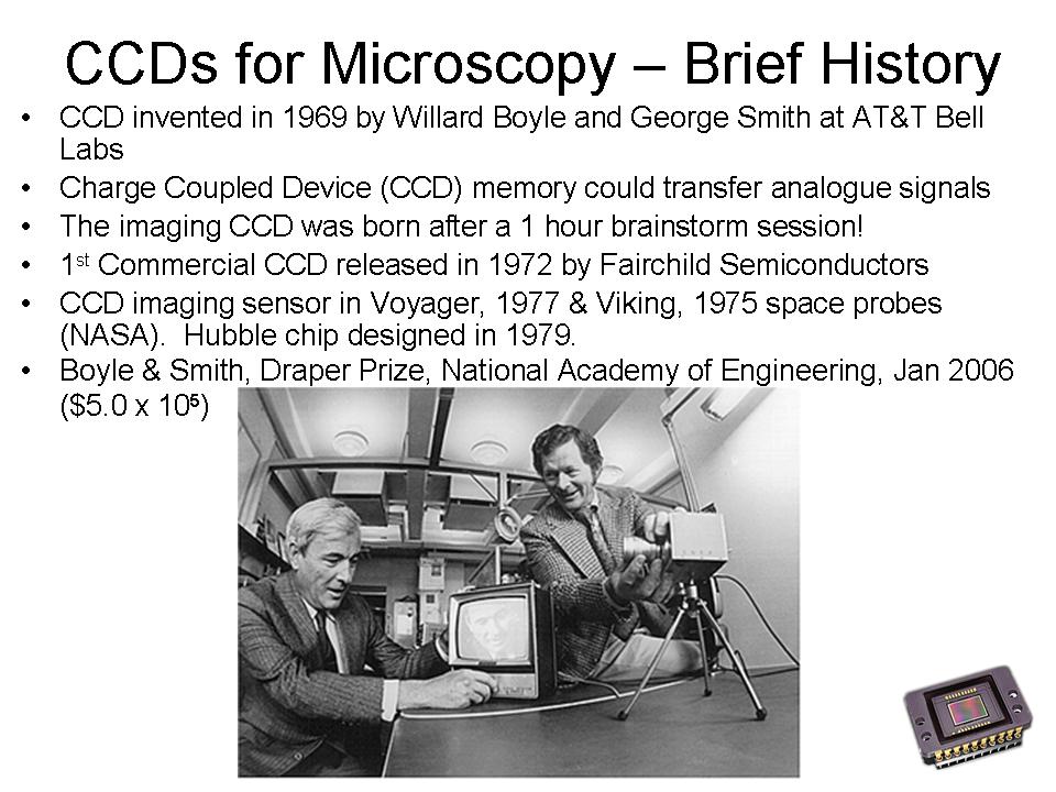 |
| Charge Coupled Device (CCD) invented in 1969 by Willard Boyle and George Smith at AT&T Bell Labs. Nobel Prize 2009. |
 |
|
| Magenta | Darkfield |
| Yellow | Confocal reflection |
| Blue | DAPI |
| Green | Fluorescence |
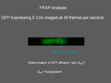
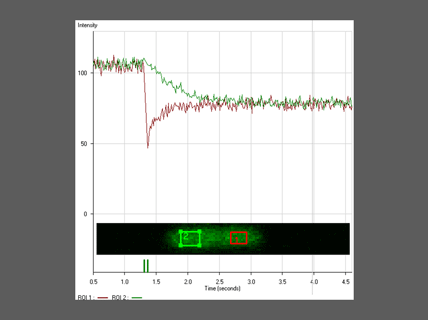
| Raster Scanning FRAP - Kristin Slade | |
 |
 |
|
|
|
|
|
|
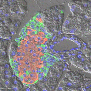
| Live Muscle Spindle - DIC with digital local contrast enhancement | Pancreatic islet - DIC + fluorescence to detect nuclei (blue), glucagon (green), and insulin (red). Dr. J. Schisler |
|
|
|
|
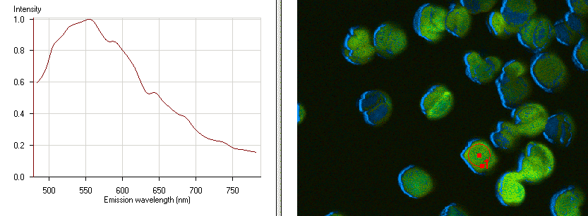
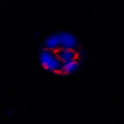
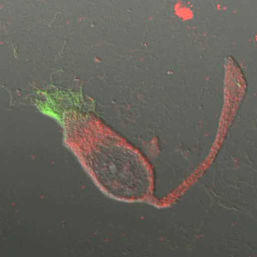
Human Red Blood Cell infected with Plasmodium falciparum |
Same view as left, showing only DB75 and Mitotracker |
|
DIC image, red Mitotracker, blue is DB75, green is DRAQ5 |
Rotation of above. Purfield, Tidwell & Mesnick |
|
|
 |
 |
 |
|
Cell membranes outlined with FM 4-64 |
Tiff format image | FM 4-64 tiff format | DIC tiff format |
|
|
|
|
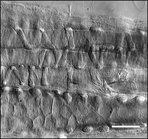
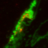
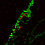
Thin transverse section of a Neuro Muscular Junction. Best wide field view of a z-series |
Same section as left deconvolved on a DeltaVisonRT imaging system |
| Red label is Alpha-bungarotoxin TexasRed. Green label is con-A FITC. Click on image to view full field of view. Sample prepared by Dr. Neal Kramarcy. Image scanned and deconvolved by M. Chua. | |
|
|
|
|
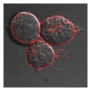
Dissociated airway epithelial cells. S. Okada et al |
Cell membranes outlined with FM 4-64. Brooke Haddock |
|
||||||||||||
|
||||||||||||
|
|
|
||||||||||
|
|
|
||||||||||
|
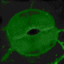
Arabidopsis guard cell labeled with GFP targeted to the membrane. Images scanned on a Zeiss 510 meta confocal microscope. Sample provided by Shouling Xu. |
||||||||||||
|
|
 |
|
| (click on image to see a larger animation) | ||
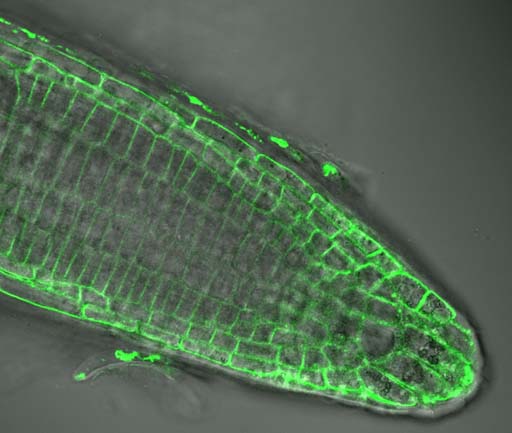
| A live root tip of Arabidopsis. Plasma membrane labeling with FM 1-43 simultaneously scanned with DIC using a Zeiss 510 confocal microscope. | Arabidopsis guard cell labeled with GFP targeted to the membrane. Rendered with Volocity 2.6.1. | Rendered with T3D |
| Please note that these images are not intended as a comparison of Volocity and T3D. Several factors including transparency and threshold are not equivalent. | ||
| Below: 3 color z-stack antibody localization in epithelial cell tissue culture, using Zeiss Meta Multitracking, with minimal intensity lasers. Image generated by Alan S Fanning, Ph.D. Cell and Molecular Physiology and Tim Oliver | |
 |
Tight Junctions are Disrupted in Epithelial Cells Expressing an Altered ZO-1 Transgene. A Madine-Darby Canine Kidney (MDCK) cell line expressing a myc-tagged ZO-1 transgene was fixed in Ethanol/Acetone and stained with antisera against the c-myc epitope tag (Cy2-green), the transmembrane protein occludin (Cy3-red), and an antiserum specific for the canine ZO-1 (Cy5-blue). The cells were imaged with a 100X PlanApo lens on the Zeiss 510 LSC Microscope using the Meta detector. Note that the transgene (red) forms large ectopic structures distinct from the normal circumferential distribution of the endogenous ZO-1 polypeptide (blue), and that the transmembrane protein occludin is also recruited into these extopic structures (green). |
| Dr. Alan Fanning | |
 |
Maximum projection of a z-series scan through a mouse embryo. Scanned on a Leica SP2 confocal microscope |
|
Mouse embryo |
|
|
Z-series scan acquired using a Zeiss 510 confocal microscope. Render using Volocity 2 made by Dr. Michael Chua Quick Time version of movie (855 KB) |
|
Arabidopsis root hair |
 |
|
|
|
| Lung airway mucus Dr. Hiro Matsui |
Fluorescent beads in fluorescent mucus |
Mucus above airway cells Dr. Ray Pickles |
Airway cilia. DIC sequence acquired at 3.9 fps using a laser scanning confocal microscope, Dr. P. Sears & Dr, A. Rossi |
 |
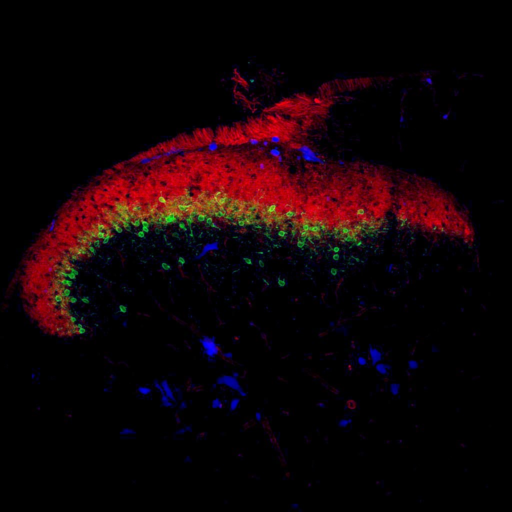 |
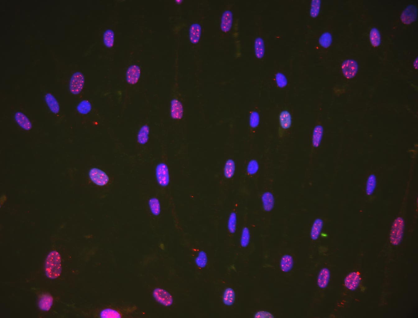
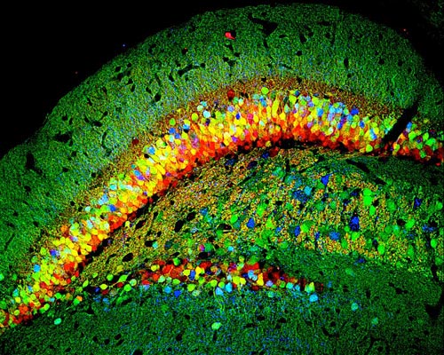
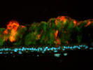
| Triple Antibody labeled sensory cortex, Dr. Sejin Huang |
Coronal section of spinal cord |
|
|
|
| Drosophila Embryo - Maximum projection of 215 sections acquired in using selective plane imaging microscopy (SPIM). Sample provided by R. Duronio Laboratory. | Mouse head CT scan - maximum projection render made with Volocity (Click image for 50 um/voxel view). Note the occlusion artifacts due to max. projection. |
 |
 |
| St. Nicolas Church, Leipzig DE. Before the Berlin Wall fell there was the Friendly Revolution of 10 June 1989 (text). | Checkpoint Charlie guard tower now at the Newseum, Washington DC |
|
|
|
|
|
|
|
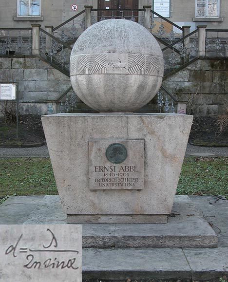
Ernst Abbe memorial at Jena, Germany: The insert, lower left, shows an enlargement of the famous diffraction equation engraved on the equator of the sphere. |
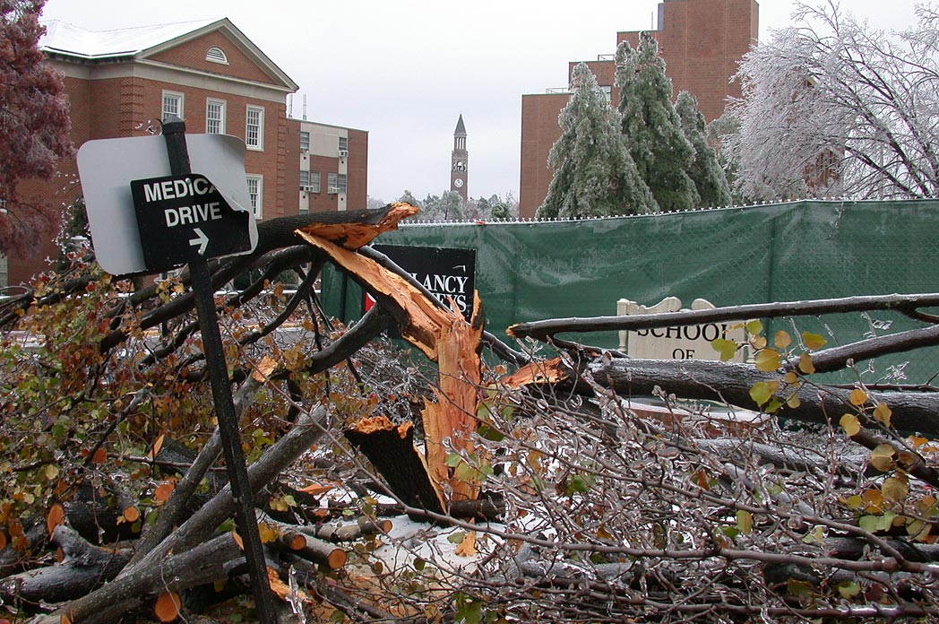
Ice storm of December 2002: Medical Drive & S. Colombia St. with the UNC Bell tower in the background. |
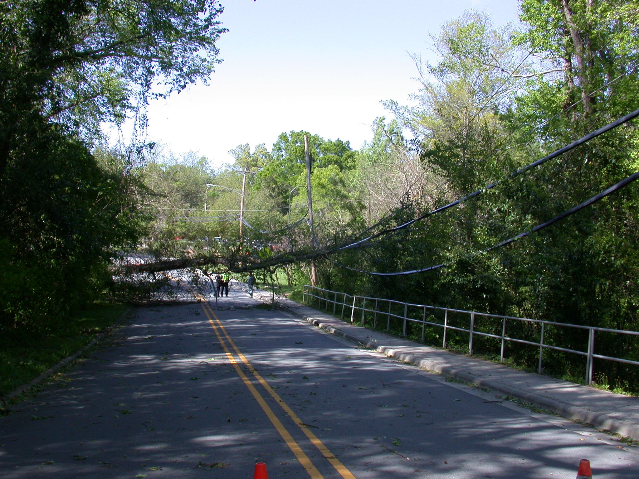
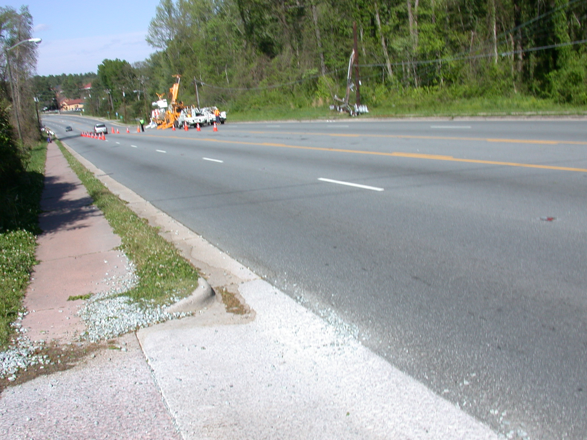
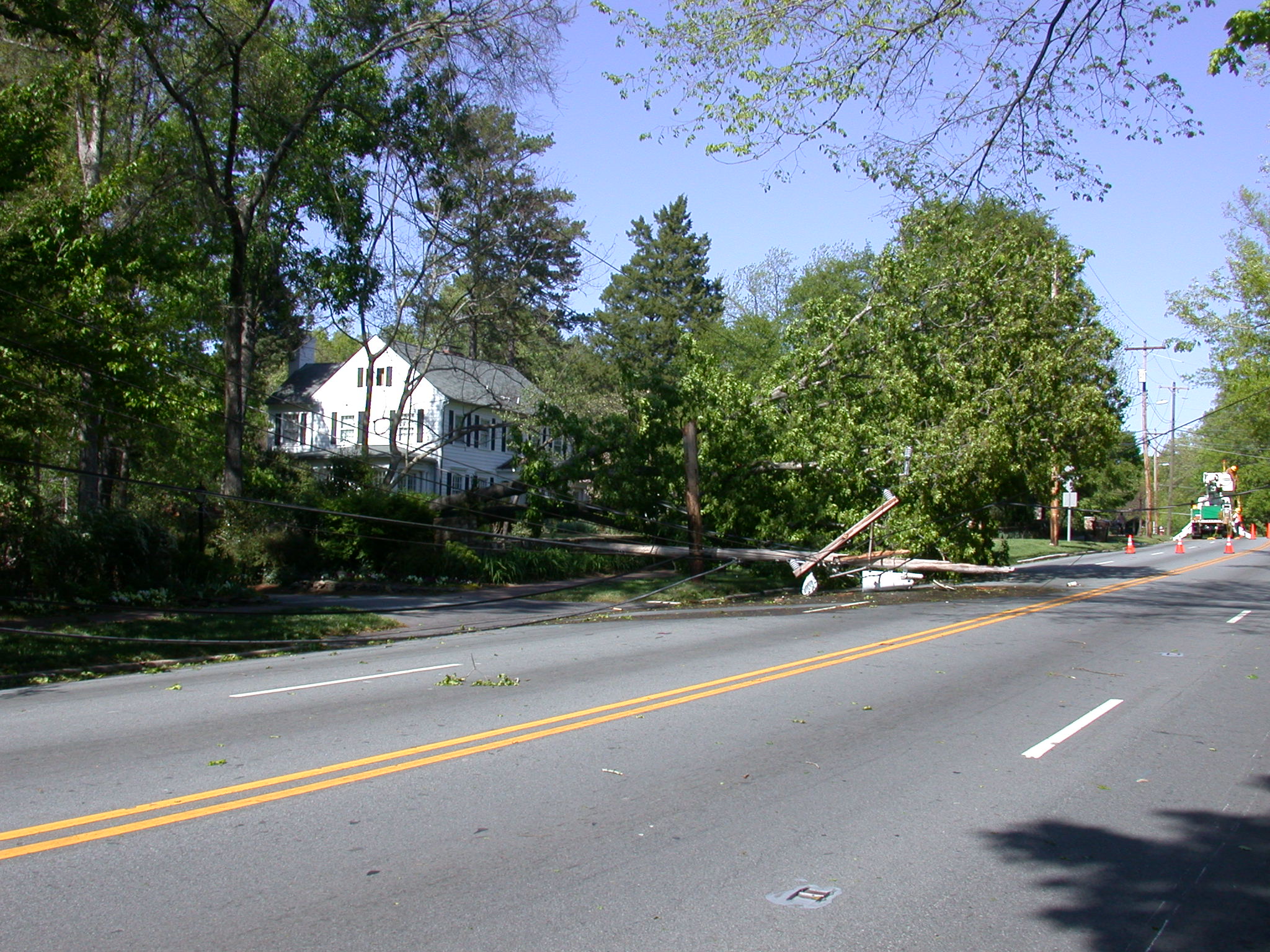
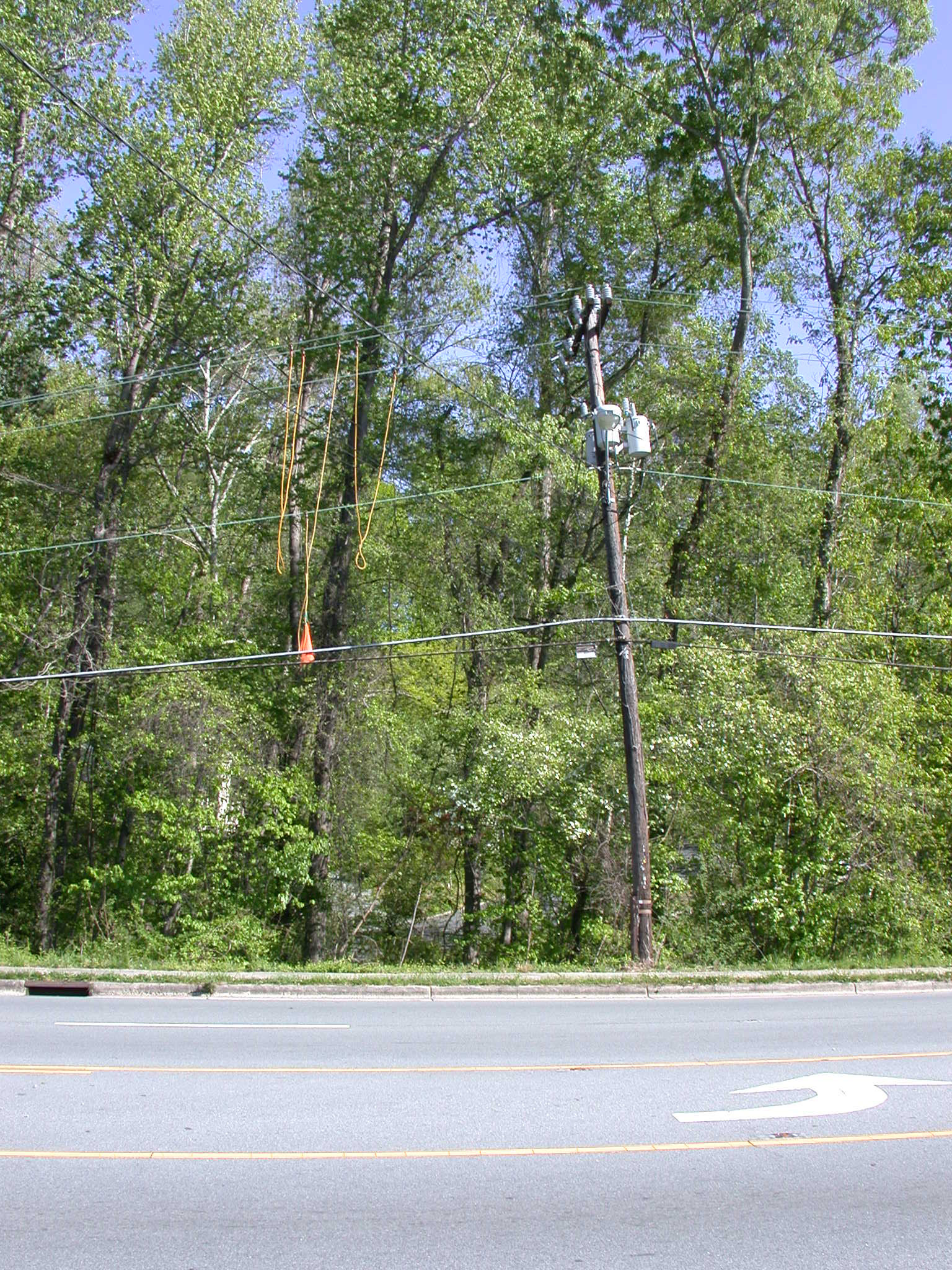
Wind Storm April 2007 |
 |
|
|
| Kitware - Medical Imaging |

 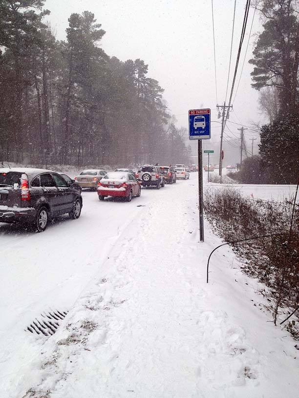 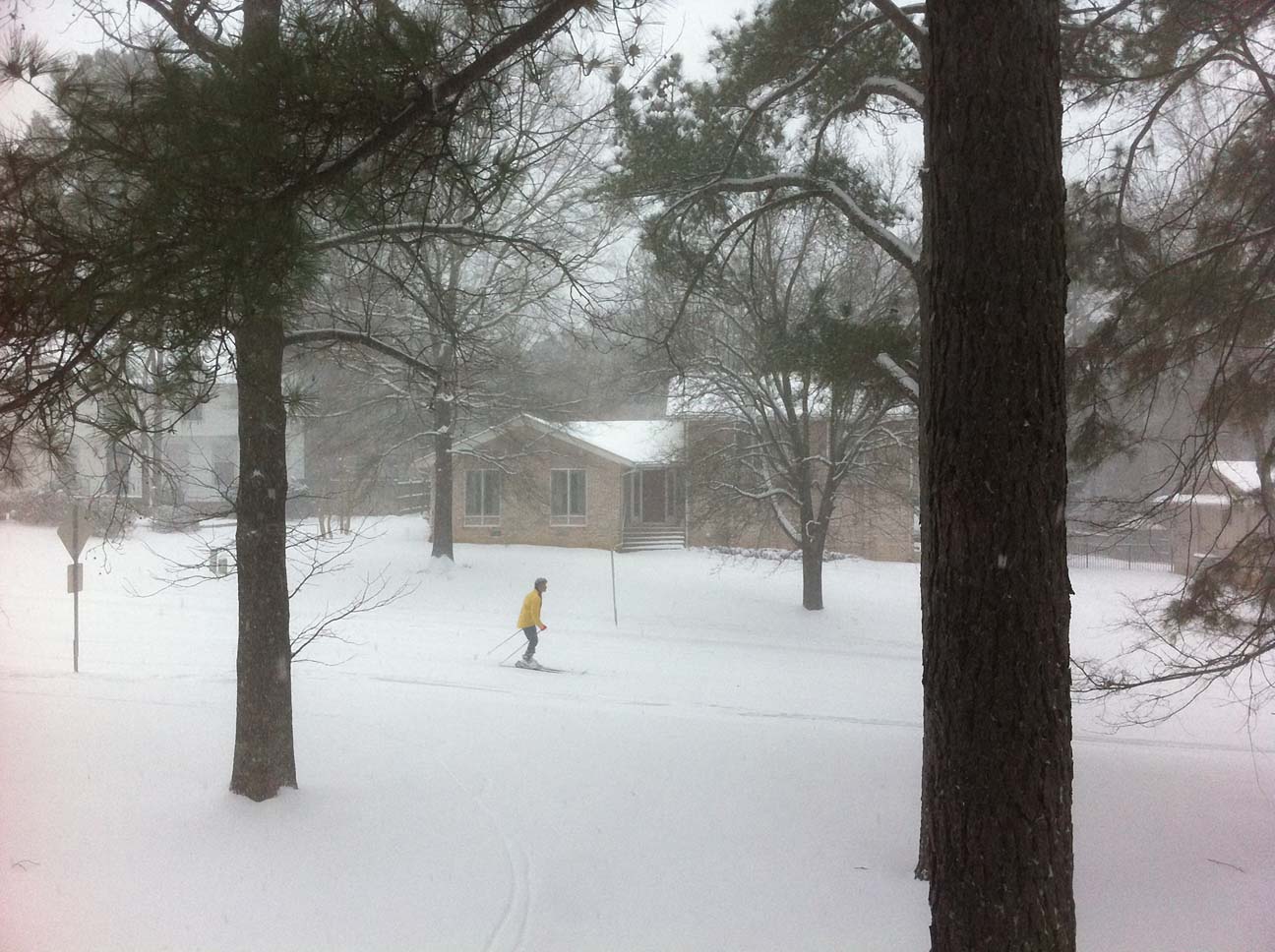 |
| Science after the snow fall - Feb 2014 |
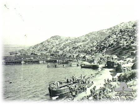
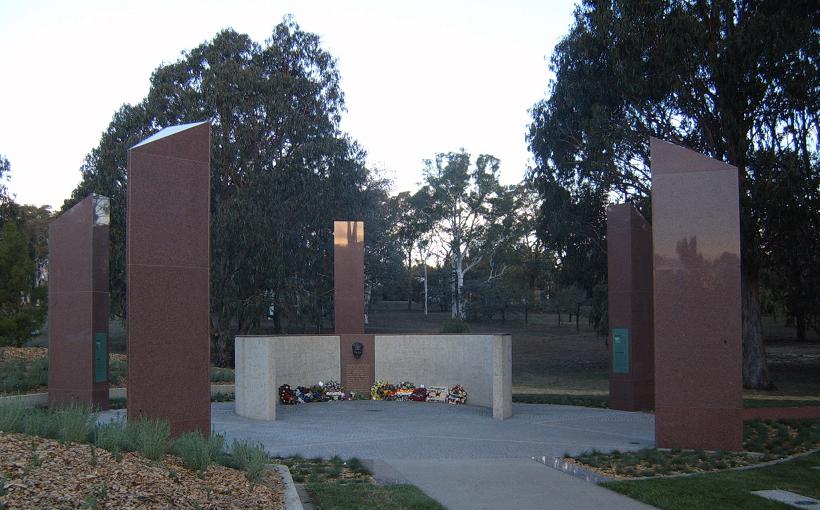 A memorial to an enemy commander on Anzac Parade. Officially the area is declared Turkish territory. |
April 25
ANZAC Day (1915) - "Those heroes that shed their blood and
lost their lives ... You are now lying in the soil of a
friendly country. Therefore rest in peace. There is no
difference between the Johnnies and the Mehmets to us where
they lie side by side here in this country of ours ... You,
the mothers who sent their sons from faraway countries, wipe
away your tears; your sons are now lying in our bosom and are
in peace. After having lost their lives on this land they have
become our sons as well." Kemal Atatürk |
 We
are saddened and shocked to hear of the passing of
Tim Oliver,
who was an employee at the Michael Hooker Microscopy
Facility before he joined
the Cell Biology Department at Duke University.
Memorial Gathering at Duke - April 15, 2011. We
are saddened and shocked to hear of the passing of
Tim Oliver,
who was an employee at the Michael Hooker Microscopy
Facility before he joined
the Cell Biology Department at Duke University.
Memorial Gathering at Duke - April 15, 2011. |
|
|
|
Copyright 2001-14 Dr. M. Chua, Office of Research, University of North Carolina, Chapel Hill, NC 27599 |
| Go Back | Booking Resources | Questions/Comments: Michael Chua |
|
|
Last Updated: 2015-08-06 |


