Michael Hooker Microscopy Facility (MHMF)
|
|
||
|
|
Michael Hooker Microscopy Facility (MHMF) |
|
|
|
||

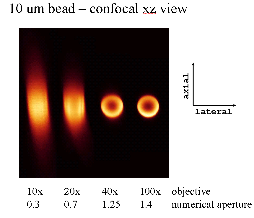
X-Z axis confocal scans of a 10 um fluorescent bead viewed with 10x dry, 20x oil, 40x oil & 100x oil objectives. The images have been scaled to display the bead at the same size. Note the loss of z-axis resolution when scanning with the lower NA objectives.
|
|
|
Copyright 2001-14 Dr. M. Chua, Office of Research, University of North Carolina, Chapel Hill, NC 27599 |
| Go Back | Booking Resources | Questions/Comments: Michael Chua |
|
counter |
Last Updated: 2014-04-02 |