Michael Hooker Microscopy Facility (MHMF)
![]()
|
|
||
|
|
Michael Hooker Microscopy Facility (MHMF) |
|
|
|
||
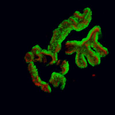
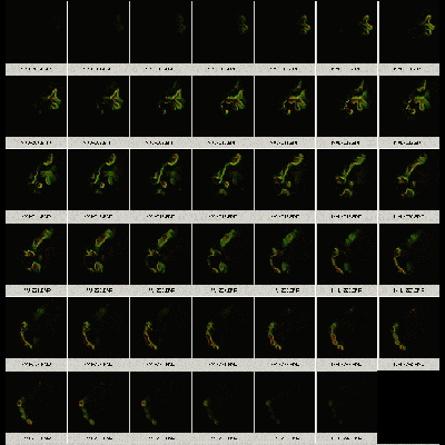
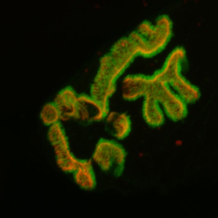
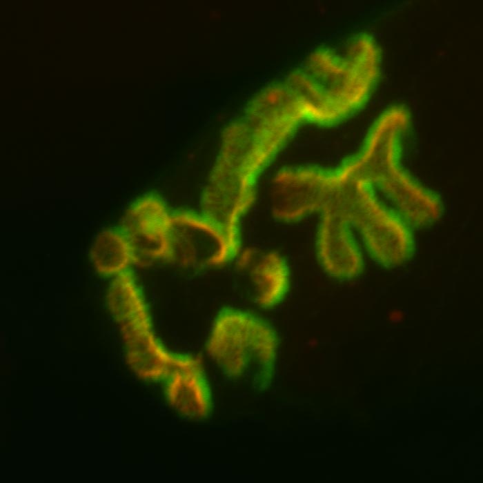
Mouse Neuromuscular Junction. Red shows receptors and green the post synaptic folds Images scanned on a Zeiss 510 meta confocal microscope. Sample provided and scanning performed by Dr. Robert Sealock.
|
|
 |
||
|
Volume render of serial optical sections using Volocity 3.0 |
Serial optical sections | ||
 |
 |
||
| Maximum projection of 41 optical sections | Average projection of 41 optical sections | ||
|
|
|
Copyright 2001-14 Dr. M. Chua, Office of Research, University of North Carolina, Chapel Hill, NC 27599 |
| Go Back | Booking Resources | Questions/Comments: Michael Chua |
|
|
Last Updated: 2010-06-22 |