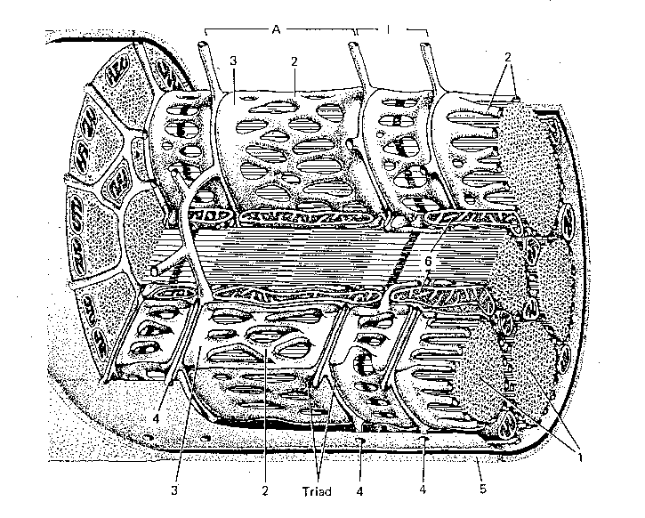

The background image shows the internal structure of a muscle fibre (cell).
Myofibrils are the protein rods which are made to slide past each other when a muscle actively contracts
Sarcoplasmic reticulum stores calcium ions and releases it to initiate contraction and pumps it in to end contraction.
Terminal cisternea are specialised regions of the sarcoplasmic reticulum which make contact with the transverse tubules. Calcium ions is released from this region onto the contractile filaments. Calcium ions trigger active sliding of the filaments, which produces muscle shortening.
Opening of the transverse tubules to the space outside of the muscle cell. Electrical signals travel from the outside surface of the muscle deep into the muscle fibre down the transverse tubules.
Myoplasm is the surface membrane of the muscle cell. This membrane carries electrical signals (the action potential) along the muscle fibre. The action potential travels into the muscle fibre down tranverse tubules.