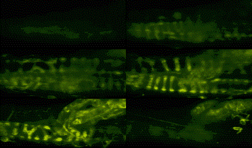
Optical Sections Through a Live Muscle Spindle

A live muscle spindle viewed as 1 um steps through the z-axis. This view was obtained with a laser scanning confocal microscope using a 40x 1.0 NA water immersion objective. The sensory nerve endings have been stained with the vitawl dye FM 1-43 (Molecular Probes). Most prominent are the annulo-spiral endings of the sensory bag nerve fibres and the myelinated axon, which clearly branch to forms an inverted y-connection with the lower bag fibre.
