Muscle Spindle Micro-Anatomy
C.C. Hunt, Dept. of Physiology, U. of North Carolina & M. Chua, Dept. of Cell Biology and Physiology, Washington U. Medical School.

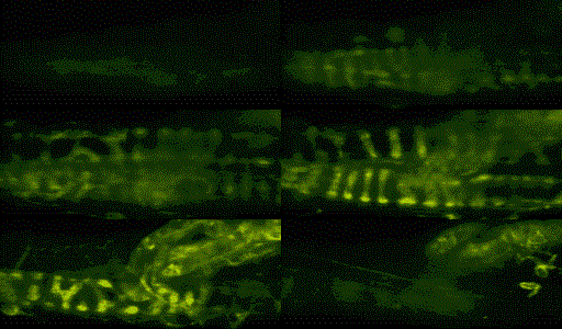
Z-section view into the sensory region of a living muscle spindle stained with FM 1-43. The larger anulo-spiral endings are around two intrfusal bag fibres. The smaller spiral endings are around intrfudal chain fibres. The sensory endings converge and leave the spindle as myelinated nerve trunks (1a afferent nerve). Z-section steps 1um.

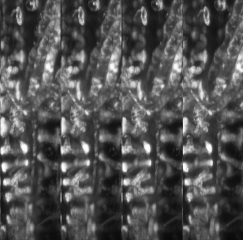
3-D reconstruction of above spindle. The stripes are views reconstructed at 10 degree rotations. A true 3-D view can be seen by observing this image at about 10" and crossing one's gaze. The sensory endings of a bag fibre are most prominent in this view.
Return to Cell Biology Confocal Home page.